Brain atrophy causes include normal aging, which starts shrinking the brain in the 30s or 40s, and disease processes such as Alzheimer’s. Exercise and adequate B vitamin status are the main natural levers, but the evidence behind each differs.
The sections below follow the sequence from baseline shrinkage to disease, then test each natural option against imaging evidence. For the wider context, see our guide to healthy aging naturally.
What causes brain atrophy as we age
Brain atrophy is the gradual loss of brain tissue. In older adults it usually reflects a normal aging baseline, with disease layered on top in some people. The stages below separate the two.
Stage one: the midlife baseline
According to WebMD’s overview of brain shrinkage, the brain’s overall size begins to shrink in your 30s or 40s, and the rate of shrinkage increases once you reach age 60. Some loss is therefore expected rather than a sign of illness. What changes the picture is pace and pattern, so a single scan result means little without a clinician’s reading of it in context.
This baseline is also why prevention advice should start earlier than retirement age. The process begins decades before most people notice any change in memory or thinking.
Stage two: uneven regional loss
Shrinkage is not uniform. The same WebMD overview notes that some areas shrink more and faster than others. A person’s overall brain size can therefore mask a lot of variation between regions. This is why researchers who study atrophy usually measure specific regions or compartments rather than relying on a single whole-brain impression.
Stage three: disease-driven loss
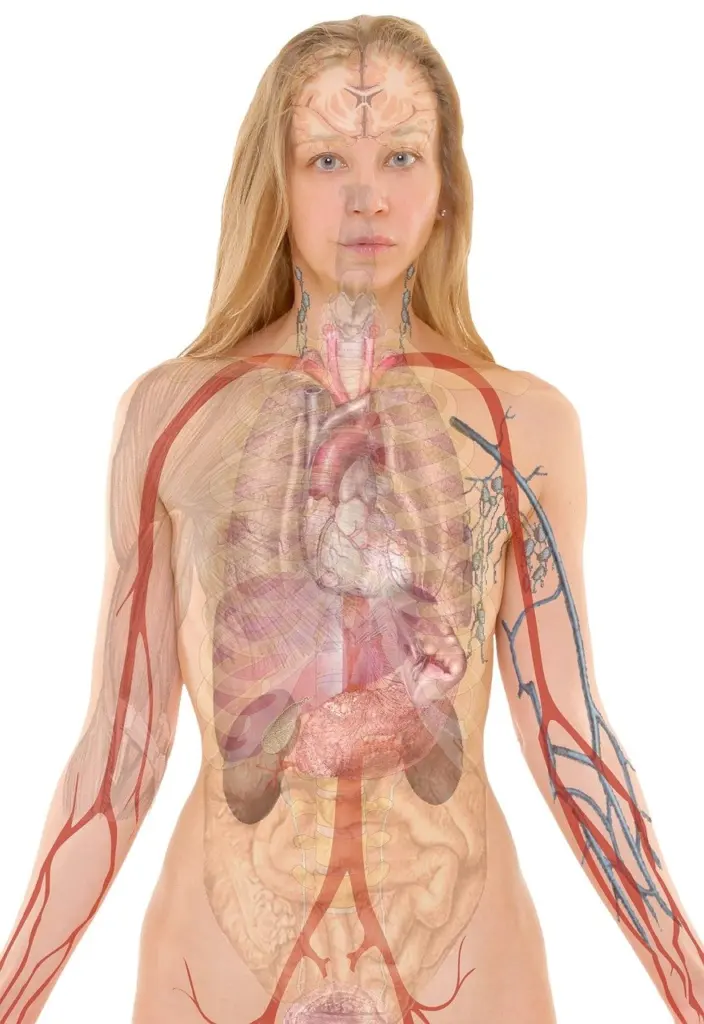
Alzheimer’s disease adds a faster, more damaging layer. The Alzheimer’s Society describes the cortex becoming thinner and the brain gradually shrinking as damage spreads. It also notes that among the areas often damaged first are the hippocampus and its connected structures. Where tissue is lost matters alongside how much, so a scan report that names the hippocampus carries different weight from a general comment about age-related change.
Practically, ordinary shrinkage is something to slow where possible, while shrinkage tied to disease needs medical assessment. Natural measures can support the first goal but are not a substitute for diagnosis in the second.
How brain volume loss is measured
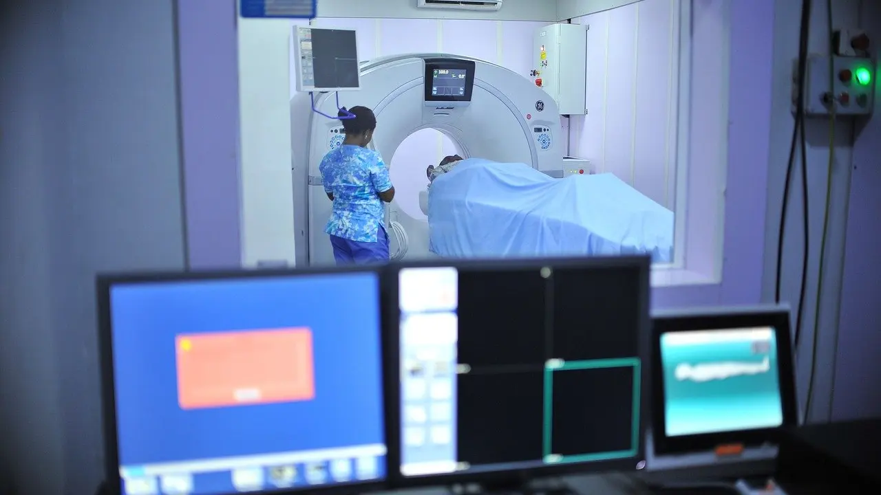
Magnetic resonance imaging (MRI) is the main tool for seeing atrophy in a living person. The Alzheimer’s Society notes that modern brain scans may show areas of reduced activity or loss of brain tissue while the person is alive. The Cleveland Clinic lists brain MRI among the tests healthcare providers may use to diagnose posterior cortical atrophy, a condition defined by tissue loss in one part of the brain.
Research uses scans in more specific ways. In a study of 36 healthy elderly people, Australian National University researchers used MRI to measure lateral ventricle-brain ratios as markers of central atrophy. They also assessed cortical atrophy and white matter hyperintensities. The ventricles are the fluid-filled spaces inside the brain, so a larger ratio of ventricle to brain points to loss of surrounding tissue.
The takeaway for readers is that “brain volume” can mean several different measurements. When comparing studies or discussing a report with a doctor, ask which region or measure is being described.
Homocysteine, B12 and folate: what the imaging shows
The clearest imaging evidence on nutrition in the supplied research links homocysteine, an amino acid in the blood, to atrophy in the centre of the brain. The table summarises the main findings from the Australian National University study.
| Measure | Finding | Planning use |
|---|---|---|
| Homocysteine and anterior ventricle-brain ratio | r = 0.49, 36 healthy elderly people | The strongest link in the study, so central atrophy is where homocysteine matters most. |
| Homocysteine and middle ventricular region | r = 0.43 | A similar but slightly weaker link, supporting the same central pattern. |
| Homocysteine and cortical atrophy or white matter hyperintensities | No association | Homocysteine is not a marker for every type of brain change. |
| Elevated homocysteine and anterior ratio of 0.34 or more, after adjustment | OR = 2.3 (95% CI 1.03-5.09) | A raised level was a significant determinant, but the wide interval means the size of the effect is uncertain. |
These figures come from the Australian National University study of plasma homocysteine and brain atrophy.
Two details strengthen the result. The analysis controlled for age, folate, B12, creatinine and white matter disease, so the homocysteine link was not simply a stand-in for those factors. Homocysteine also did not track every imaging measure, which suggests a specific relationship with central atrophy rather than general brain ill health.
The limits are just as important. This is an observational analysis of 36 people, so it shows that higher homocysteine goes with more central atrophy, not that lowering homocysteine reverses it. The lower end of the confidence interval sits close to no effect, so treat the result as a signal worth acting on cautiously rather than as proof.
B vitamins and brain shrinkage: what to do
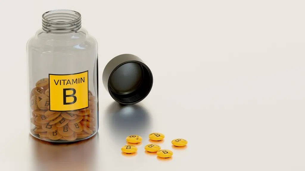
B vitamins are the best-supported nutritional lever in this evidence, with a clear condition: they matter most when intake or status is low. WebMD reports that without vitamin B, older adults are at greater risk of brain shrinkage and cognitive decline. Combined with the homocysteine findings above, the logic is consistent. Low B12 and folate status goes with higher homocysteine, and higher homocysteine goes with more central atrophy.

What this means in practice:
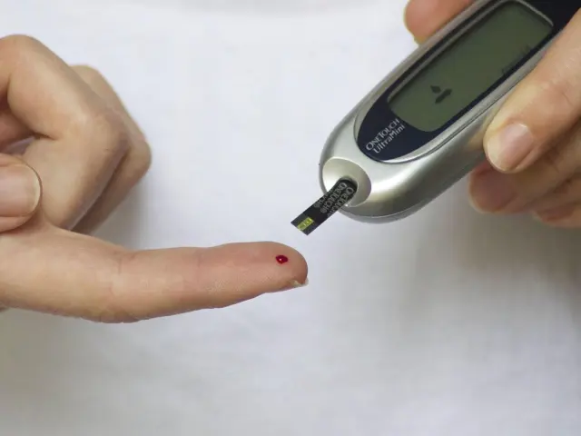
- Check status before supplementing. The evidence ties shrinkage risk to low vitamin B, so people with possible deficiency gain the most from knowing their levels. A clinician can arrange B12 and folate testing.
- Do not assume more is better. The imaging study adjusted for folate and B12 and still found a homocysteine effect, and nothing supplied shows that high-dose supplements slow shrinkage in people with adequate levels.
- Treat supplements as a correction, not a cure. If a deficiency is found, correcting it removes one plausible contributor to atrophy. It does not address the other stages described earlier.
Anyone who is already experiencing memory or thinking changes should raise them with a doctor rather than relying on a vitamin to deal with them.
Exercise and brain volume
Exercise is the other main lever, and the supported mechanism is blood supply. WebMD states that getting exercise increases blood flow to your entire body, especially your brain. Since brain tissue depends on that supply, regular aerobic activity is a reasonable foundation for protecting the brain as it ages.
The honest answer to whether exercise increases brain volume is more measured. The supplied evidence supports increased blood flow to the brain, not a measured gain in tissue. Think of exercise as a way to support the brain and slow decline rather than to rebuild what is lost. That framing also suits the timeline: shrinkage starts in midlife and accelerates from age 60, so activity pays off most when it is a long-standing habit.
Choose an activity you can sustain, because consistency matters more than intensity for a benefit built over decades. Walking suits most people. Anyone with a heart condition or mobility limits should agree a plan with their doctor first.

Putting the evidence together
The research is strongest on why shrinkage happens and weaker on how much natural measures can change it. A sensible order of priorities follows from that.
- Confirm your B12 and folate status. It is the only lever here with direct imaging evidence behind it, and a deficiency is simple to identify.
- Build regular aerobic activity into the week. It addresses blood flow to the brain and has no downside for most people when introduced gradually.
- Get new cognitive symptoms assessed. Disease-related loss, including the hippocampal damage seen in Alzheimer’s, needs medical evaluation that lifestyle changes cannot replace.
Brain shrinkage is one of several aging processes that interact. Cellular ageing is covered in our guide to telomeres and aging, and if you are wondering does fasting slow aging, that question has its own page.
Frequently asked questions
How much brain volume loss is normal?
Some loss is normal. It begins in the 30s or 40s and speeds up from age 60, according to WebMD. Regions shrink at different rates, so a clinician reads a scan by region and context rather than against a single cut-off.
Does exercise increase or preserve brain volume?
The supported effect is more blood flow to the brain, not a proven gain in tissue. Regular exercise is best viewed as a habit that helps preserve brain health over many years.
Is high homocysteine the same as brain atrophy?
No. Higher homocysteine was associated with atrophy in central brain regions in a study of healthy elderly people, but not with cortical atrophy or white matter hyperintensities. It is a marker linked to one pattern of loss, not a diagnosis.
Can a scan show brain atrophy in a living person?
Yes. Brain scans such as MRI can show loss of brain tissue during life, according to the Alzheimer’s Society. Interpreting the result still requires a clinician who can place it in context.